
What Does the Heart Look Like and How Does It Work?

This kind of monitoring does not constitute a complete ECG.A heart monitor only measures the rate and rhythm of the heartbeat.By comparison, a heart monitor requires only three electrode leads – one each on the right arm, left arm, and left chest.

The printed view of these recordings is the electrocardiogram.The signals received from each electrode are recorded.An electrode lead, or patch, is placed on each arm and leg and six are placed across the chest wall.Ten electrodes are needed to produce 12 electrical views of the heart.A standardized system has been developed for the electrode placement for a routine ECG.The electrocardiogram can measure the rate and rhythm of the heartbeat, as well as provide indirect evidence of blood flow to the heart muscle.The heart is a two stage electrical pump and the heart's electrical activity can be measured by electrodes placed on the skin.Numerous textbooks are devoted to the subject. While it is a relatively simple test to perform, the interpretation of the ECG tracing requires significant amounts of training. The electrocardiogram (ECG or EKG) is a diagnostic tool that is routinely used to assess the electrical and muscular functions of the heart. S2CID 6895831.Picture of the basic anatomy of the heart "A Simple Ballistocardiographic System For A Medical Cardiovascular Physiology Course". Archived from the original on 8 February 2007. Simultaneous Monitoring of Ballistocardiogram and Photoplethysmogram Using a Camera Dangdang Shao, "IEEE Transactions on Biomedical Engineering", Volume: 64, Issue: 5, May 2017, p. 1003–1010.Half a century of contributing to medical care and society.IEEE Transactions on Biomedical Engineering. "Simultaneous Monitoring of Ballistocardiogram and Photoplethysmogram Using a Camera". ^ Shao, Dangdang Tsow, Francis Liu, Chenbin Yang, Yuting Tao, Nongjian (2017)."Theory and Developments in an Unobtrusive Cardiovascular System Representation: Ballistocardiography". ^ "Ballistocardiography, a bibliography"."Ballistocardiogram: Mechanism and Potential for Unobtrusive Cardiovascular Health Monitoring". "Certain Molar Movements of the Human Body produced by the Circulation of the Blood". National Library of Medicine Medical Subject Headings (MeSH)
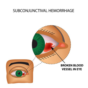
#Heart blood vessel broken cardiograph image plus
The term ballistocardiograph originated from the Roman ballista, which is derived from the Greek word ballein (to throw), a machine for launching missiles, plus the Greek words for heart and writing. A BCG scale is able to show a person's heart rate as well as their weight. One example of the use of a BCG is a ballistocardiographic scale, which measures the recoil of the persons body who is on the scale. Recent work also validates BCG could be monitored using camera in a non-contact manner. Isaac Starr, that the effect of main heart malfunctions can be identified by observing and analyzing the BCG signal. It was shown for the first time, after an extensive research work by Dr. It is a vital sign in the 1–20 Hz frequency range which is caused by the mechanical movement of the heart and can be recorded by noninvasive methods from the surface of the body. Ballistocardiography is a technique for producing a graphical representation of repetitive motions of the human body arising from the sudden ejection of blood into the great vessels with each heart beat. As different parts of the aorta expand and contract, the body continues to move downward and upward in a repeating pattern. The downward movement of blood through the descending aorta produces an upward recoil, moving the body upward with each heartbeat. The ballistocardiograph ( BCG) is a measure of ballistic forces generated by the heart.


 0 kommentar(er)
